Abstract
Objective: Myelodysplastic syndrome (MDS) is a group of heterogeneous hematopoietic clonal diseases. Recent studies have confirmed that there are essential immune differences in the progression of MDS. However, the detailed mechanisms remain unclear. In order to explore the changes of immune disorders during the occurrence and progression of MDS, and identify critical differentially expressed factors as well as prognostic relevance. What's more, to probe the possible immunological mechanism in the progression of MDS.
Methods: The study had a prospective design. A total of 86 patients with newly diagnosed MDS were enrolled in this study, where patients were classified as low risk group and high risk group based on the revised international prognostic scoring system (IPSS-R) score. The expression profiles of multiple cytokines in patients’ plasma were determined using Luminex microarray assay, and flow cytometry were used to measure lymphocyte subsets in peripheral blood, especially T regulatory cell (Treg) and its subsets: induced regulatory Treg (iTreg: CD45RO+CD3+CD4+CD25+CD127low+) and naïve regulated Treg (nTreg: CD45RA+CD3+CD4+CD25+CD127low+), and the percentage of Monocytic-myeloid derived suppressor cells (M-MDSCs) in bone marrow. The mRNA expression of NLRP3 inflammasome related molecules was detected by real-time quantitative PCR. P <0 .05 was considered statistically significant.
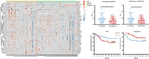
Results: In the high risk group, the plasma levels of some cytokines or chemokines with promoting or amplifying inflammation signals in directly or indirectly way, including IL-9, MIP-1β,GRO-α and TNF-β, and two growth factors influencing the immune microenvironment by regulating the growth and differentiation of corresponding cells, including β-NGF, PDGF-bb, were significantly lower (P<0.05).The expression level of IL-1β was significantly higher in the high risk group (P<0.05) and its expression was significantly correlated with the percentage of M-MDSCs (P<0.0001). What's more, the percentage of bone marrow M-MDSCs facilitated immune tolerance was significantly higher. Although there is no significant difference of Treg between the two groups, both the percentages and absolute counts of nTreg/iTreg was decreased significantly in high-risk group of MDS patients (P<0.05) (Figure). After adjusting the Cox model, nTreg/iTreg was an independent protective factor for OS (P=0.038, HR=0.081, 95% CI: 0.008-0.869), and elevated IL-1β level was an independent prognostic factor for PFS (P=0.018, HR=2.554, 95% CI: 1.175-5.552). Then, we found that the mRNA expression of NLRP3, ASC, Caspase 1 and IL-1β were significantly increased in the high risk group(P<0.05).
Conclusion: All above results demonstrated that the expression of pro-inflammatory cytokines and chemokines was increased in low risk MDS, while the amplification of bone marrow M-MDSCs and the transformation of nTreg to iTreg may be the key immune mechanisms leading to disease progression. The activation of NLRP3/IL-1β pathway may play an important role in the progression of disease. The serum levels of IL-1β and the percentage of nTreg /iTreg ratio may be an effective indicator to predict the prognosis of MDS. It provided a basis for clinical prediction and early intervention to improve the prognosis.
Disclosures
No relevant conflicts of interest to declare.
Author notes
Asterisk with author names denotes non-ASH members.


This feature is available to Subscribers Only
Sign In or Create an Account Close Modal